CBCT Imaging in General Dental Practice: When It's Worth the Investment | WSS
Practice Operations
Practice Operations · Western Surgical and Sedation
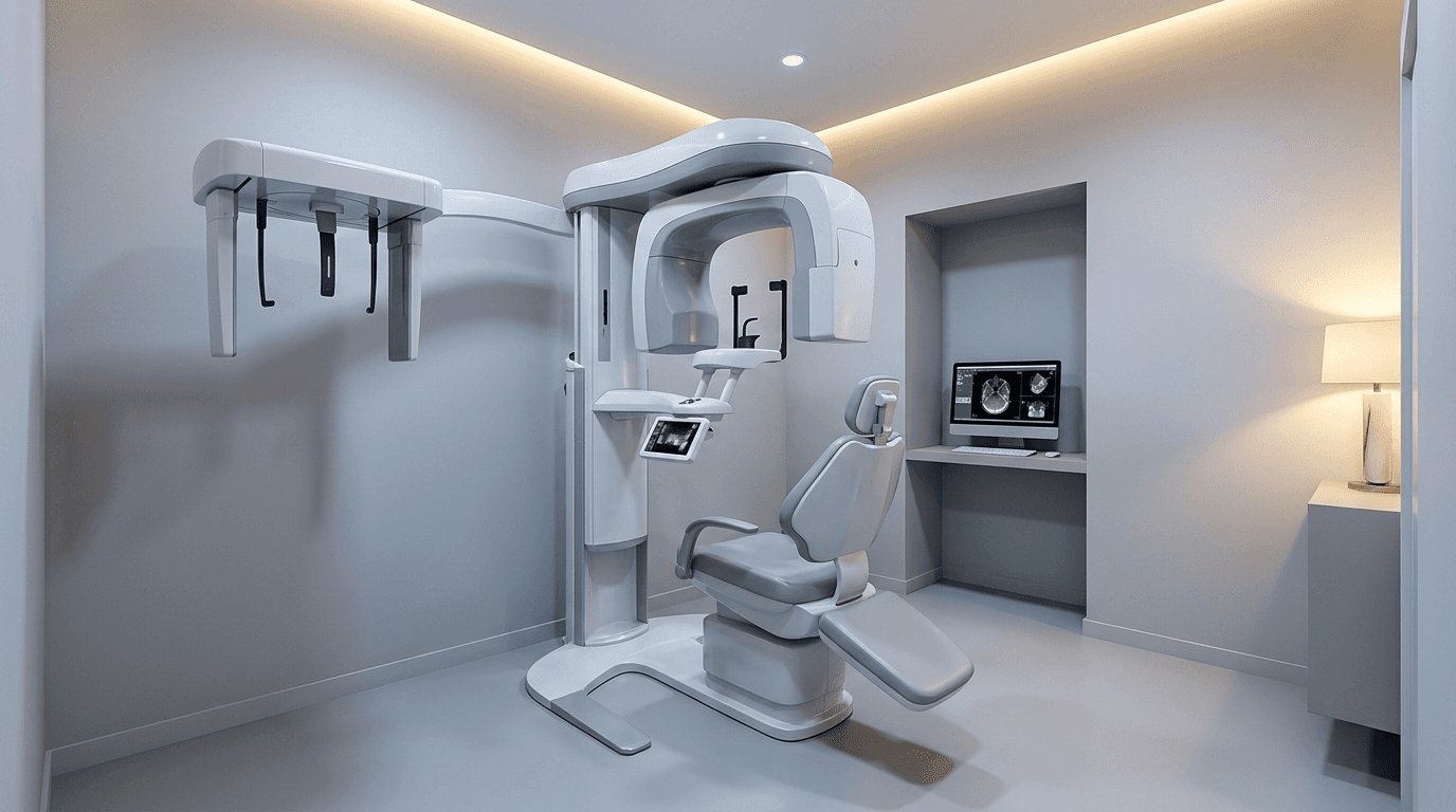
Cone beam computed tomography has moved from a specialist technology to something increasingly common in general dental practice. The clinical value is well established — three-dimensional imaging reveals anatomy that two-dimensional radiographs cannot show, and that additional information changes treatment decisions in specific, identifiable situations.
The question for most general dentists is not whether CBCT provides clinical value. It clearly does. The question is whether the volume of cases in their specific practice justifies the investment, or whether a referral relationship for imaging serves the practice better.
This article addresses that decision directly: where CBCT genuinely changes clinical decisions, what the realistic cost picture looks like, and how expanding surgical scope affects the calculation.
What CBCT Shows That Panoramic Imaging Cannot
A panoramic radiograph compresses three-dimensional anatomy into a two-dimensional image. This works well for many diagnostic purposes but introduces specific limitations that matter significantly in certain clinical situations.
Buccolingual position
A panoramic radiograph shows vertical and horizontal relationships but cannot show whether a structure is positioned toward the cheek or the tongue. For an impacted third molar sitting near the inferior alveolar nerve, this dimension determines whether the roots are actually in contact with the nerve canal or merely appear superimposed on it in two dimensions.
True bone volume and density
For implant planning, understanding available bone width — not just height — is essential. A site that appears to have adequate bone on a panoramic view may have a narrow ridge that cannot accommodate an implant without grafting. CBCT reveals this before the surgical appointment rather than during it.
Pathology characterization
Cysts, lesions, and other pathology are more accurately localized and characterized in three dimensions, informing both the treatment plan and the referral decision when specialist involvement is appropriate.
Root morphology
Complex root anatomy, including dilaceration, additional canals, and unusual configurations, is more accurately assessed with three-dimensional imaging — relevant both for endodontic treatment and for surgical extraction planning.
When CBCT Actually Changes the Clinical Decision
This is the practical filter for evaluating CBCT value. Imaging that produces more information but does not change what you do has limited clinical value. Imaging that changes the treatment plan, the surgical approach, or the referral decision has clear value.

Third molar extraction with suspected nerve proximity
When panoramic imaging shows signs of significant inferior alveolar nerve proximity — root darkening, canal deflection, loss of the canal's cortical outline — CBCT determines whether there is true contact. This changes the surgical approach, may indicate coronectomy rather than complete extraction, and substantially changes the informed consent conversation.
Implant site assessment
For any implant case, CBCT determines whether the site has adequate bone volume for placement, whether grafting is required, and where the implant should be positioned relative to anatomical structures. This is now considered standard of care for implant planning in most contexts.
Complex or atypical anatomy
Cases where clinical or panoramic findings suggest something unusual — an unexpected radiolucency, an atypical eruption pattern, suspected pathology — benefit from three-dimensional characterization before proceeding.
The clearest indication for CBCT is a specific clinical question that two-dimensional imaging cannot answer and that will change what you do. Imaging without that question produces radiation exposure and cost without corresponding clinical benefit.
The Cost Picture
An honest evaluation of CBCT investment requires accounting for more than the equipment purchase price.
Capital cost
CBCT units for dental practice range broadly in price depending on field of view, image quality, and included software. This is a significant capital investment typically financed over several years.
Ongoing costs
Software licensing and updates for planning and viewing software
Service contracts and maintenance
Staff training on operation and safety protocols
Radiation safety compliance, including state registration and periodic inspection depending on jurisdiction
Space allocation — CBCT requires a dedicated area meeting shielding requirements
The referral alternative
Practices that do not own CBCT typically establish a relationship with a local imaging center or specialist practice that provides scans on referral. This introduces a scheduling step and patient inconvenience but eliminates capital cost entirely. For practices with low CBCT case volume, this is frequently the more economical approach.
How Surgical Scope Changes the Calculation
The CBCT investment calculation shifts significantly for practices that have expanded into surgical procedures — specifically third molar extraction and implant placement.
A general practice that refers all surgical cases has relatively few situations where CBCT changes a decision. A practice performing regular third molar extractions and implant placements has a fundamentally different case mix, with CBCT indicated in a meaningful percentage of cases.
A practical volume threshold
Practices should estimate their monthly volume of cases where CBCT would genuinely change a clinical decision — nerve-proximity third molars, implant planning cases, and complex pathology. When this volume reaches a level where referral scheduling becomes a recurring friction point and the imaging fees paid to outside providers become substantial, in-house CBCT becomes worth serious evaluation.
Below that threshold, a strong referral relationship with a local imaging provider serves the practice well without the capital commitment.
Radiation Safety and Appropriate Use
CBCT delivers higher radiation dose than conventional dental radiography, and appropriate use guidelines emphasize that the imaging should be justified by a specific clinical question rather than obtained routinely.
Professional guidelines consistently recommend that CBCT be used when the diagnostic information it provides cannot be obtained from lower-dose imaging and when that information will affect patient management. Practices should have a documented protocol establishing the clinical indications for CBCT in their practice, and each scan should be justifiable against that protocol.
Field of view selection also matters — using the smallest field of view that answers the clinical question reduces patient dose while providing the necessary diagnostic information.
Frequently Asked Questions
Is CBCT required for all third molar extractions?
No. CBCT is indicated when panoramic imaging shows specific signs of significant inferior alveolar nerve proximity or other anatomical complexity. Routine CBCT for all third molar cases is not supported by current appropriate-use guidelines and adds unnecessary radiation exposure and cost.
Can I bill separately for CBCT imaging?
CBCT has specific procedure codes and is billable separately from other procedures performed at the same visit. Coverage varies by plan and often depends on documented clinical indication, so benefits verification is advisable for cases where CBCT is planned in advance.
What field of view should a general practice purchase?
This depends on intended clinical use. Smaller fields of view are adequate for single-site implant planning and localized third molar assessment while delivering lower patient dose. Larger fields of view support broader diagnostic applications but at higher dose and typically higher equipment cost. Most general practices are well served by units offering selectable field of view.
How long does it take to become proficient at reading CBCT scans?
Basic proficiency for the specific applications a general practice uses most — implant site assessment and nerve proximity evaluation — develops relatively quickly with structured training and repetition. Comprehensive interpretation of full-volume scans, including incidental findings outside the area of clinical interest, requires more extensive training. Many practices establish a relationship with an oral and maxillofacial radiologist for formal interpretation of full-volume scans.
Technology supports technique. It doesn't replace it.
Western Surgical and Sedation trains general dentists in the surgical judgment and technique that make advanced imaging genuinely useful — starting with live patient experience.
Explore our programs at westernsurgicalandsedation.com/courses






Related Articles
Related reading
Explore articles connected to surgical training, IV moderate sedation, and real world clinical decision making, selected to support dentists applying advanced care in daily practice.